
Introduction
A research centre, founded with money from Arthritis Research UK(1), provided a grant to support gruesome and protracted experiments, which were conducted on a number of ten-week-old, male mice. The experiments involved operating on the mice to intentionally damage one knee of each animal. Animals previously mutilated in this way had been killed over a period of time (between two to eight weeks after the operation) and the injured joint sliced, labelled with a substance called 1-11E, or a control substance, and examined.
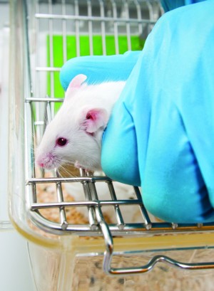
More mice were surgically mutilated as described above. They received injections, either into their damaged knee or a vein, four weeks and eight weeks after the surgery. Following the injections, ‘live’ images were taken of the animals’ knees, for up to a fortnight. After the final imaging, the mice were killed by having their necks broken (2).
The aim of the experiments was to test whether 1-11E could be used to monitor early disease in the animal model of osteoarthritis.
Animal Suffering
- Following the operation to damage their knees, but before they recovered from the anaesthetic, the mice were given a painkilling injection. No information is provided about the dose given, so it is impossible to determine whether this would have provided adequate pain relief.
- The mice were ‘monitored daily post-surgery’, which is wholly inadequate. At this frequency, mice who were in pain or otherwise required urgent attention may not have been discovered for many hours. To only monitor animals once a day is insufficient even for checking everyday elements—such as the animals’ water, whether there has been any fighting, etc. It is certainly unacceptable for animals who have recently undergone surgery.
- It has been acknowledged that there are differences in cartilage thickness and volume between boys and girls and in the prevalence, incidence and severity of osteoarthritis (OA) between men and women (3). The difference in the severity of OA between male and female mice has also been stated in a paper concerning surgical models of OA (4), with the severity in males being higher. The experiment used only male mice; there is no explanation of why this was done.
- The mice would frequently be caught and either sedated or anaesthetised in order to image their joints. Capture, restraint and injections would be stressful for the animals – there is no mention of the mice being made more familiar with these procedures, in an attempt to lessen the distress caused.
- A paper which reported the same surgery in other groups of mice stated that this type of surgery can cause moderate to severe ‘lesions’ to the cartilage in the joints of mice within 8 weeks of the operation. (5)
The experiments
- A compound called 1-11E was investigated as a possible way to mark early OA disease so it could be imaged (‘seen’ inside the body).
- Preserved samples from the knees of one group of mice were treated with 1-11E or another molecule, C7, both of which were fused to a dye. The C7 was to act as a control. The sections of damaged knee were examined to see how the 1-11E had stained the damaged tissue.
- Ten-week-old male mice were operated on, their knee joint opened and a ligament cut in order to destabilise their joint (this was the ‘DMM’ group). Another group of mice had their knee joint opened and closed again (this was the ‘sham’ group).
- Four and eight weeks after DMM surgery, one group of mice were injected, into their damaged knee, with one of the two compounds described above. The animals’ knees were shaved to enable the dye to be seen within their bodies. Images were taken at different time points up to two weeks after the injection into the knee.
- Four and eight weeks after DMM surgery, another group of the mice were injected, intravenously, with one of two compounds similar to those described above. Images were taken of the animals’ knees 4, 8, 24 and 48 hours after the injection.
- When the animals had been imaged for the final time they were killed by having their necks broken.
Faulty science - A 2010 paper (6) explains the development of 1-11E and how, with human cartilage, it bound only to damaged cartilage, characteristic of arthritis, not normal cartilage. The paper concludes that 1-11E could deliver a substance specifically to the affected joint, ‘even when applied systemically,’ i.e. when it was injected into the mice’s abdomens.
- The conclusion of the 2015 paper was that 1-11E binds specifically to early OA cartilage and can be visualised after being injected into the knee or a vein. It was already known from the 2010 paper that 1-11E accumulates in arthritic joints, even if not injected there.
- There are differences in how OA affects male or female mice, mice of different ages and also men and women. The extrapolation of results from mice to humans is unreliable. This is likely to be even more relevant when considering that the researchers have artificially induced disease in relatively young animals whereas osteoarthritis is a disease usually found in older people.
- A review of the numerous animal ‘models’ of osteoarthritis of the knee highlights a structural difference between the mouse knee and human knee (8).
- The research used unevenly sized experimental groups. The live imaging groups who received an injection into their damaged knee contained a different number of animals: seven animals received 1-11E and five animals received C7. The authors do not explain why the groups were uneven.
- The in vivo group, after receiving injections in their damaged knees, were not imaged the same number of times. From graphs in the paper, the 1-11E group appear to have been imaged 10 times and the control group, C7, appear to have been imaged 9 times. There is no explanation for this discrepancy.
- A paper about osteoarthritis states that it is a process which takes place over decades and is a complex interplay between many factors. (7) An operation to deliberately mutilate young, male animals followed by eight weeks of monitoring cannot ever hope to capture the numerous factors which are at play in men and women with osteoarthritis.
Additional information and background notes
- Osteoarthritis is the most common type of arthritis. It most often develops in adults who are in their late 40s or older. It is more common in women and also those with a family history of the condition. It can occur after injury or in association with other joint-related conditions. (9) Osteoarthritis affects the smooth cartilage in the joint, which leads to pain and stiffness, and movement is made difficult. When the cartilage is roughened, the tendons and ligaments must work harder, which can cause swelling and bony formations.
- In the UK, around 8 million people have osteoarthritis.
- There is no cure for osteoarthritis. Treatments available include painkillers or non-steroidal anti-inflammatories. In severe cases, joints may be replaced or fused or bones may be cut and aligned.
References
- Arthritis Research UK (ARUK) Centre for OA Pathogenesis, grant ref. 20205.
- Lim, N.H, Vincent, T.L. & Nissim, A. (2015) ‘In vivo optical imaging of early osteoarthritis using an antibody specific to damaged arthritic cartilage’. Arthritis Research & Therapy, DOI 10.1186/s13075-015-0898-5.
- Maleki-Fischbach, M. & Jordan, J.M. (2010) ‘New developments in osteoarthritis. Sex differences in magnetic resonance imaging-based biomarkers and in those of joint metabolism’, Arthritis Research & Therapy, vol.12, pp.212-221.
- Ma, H.L. et al (2006) ‘Oesteoarthritis severity is sex dependent in a surgical mouse model’, OsteoArthritis and Cartilage, vol.15, pp.695-700.
- McNulty, M.A. et al (2012) ‘Histopathology of Naturally Occurring and Surgically Induced Osteoarthritis in Mice’, Osteoarthritis Cartilage, vol.20, no.8, pp.949-956. doi: 10.1016/j.joca.2012.05.001
- Hughes, C et al (2010) ‘Human Single-Chain Variable Fragment That Specifically Targets Arthritic Cartilage’, Arthritis & Rheumatism, vol.62, no.4, pp.1007-1016.
- Chu et al (2012) ‘Early diagnosis to enable early treatment of pre-osteoarthritis’, Arthritis Research & Therapy, vol.14, pp.212-221.
- Gregory, M.H. et al (2012) ‘A review of translational animal models for knee osteoarthritis.’, Arthritis, doi: 10.1155/2012/764621
- http://www.nhs.uk/Conditions/Arthritis/Pages/Introduction.aspx