
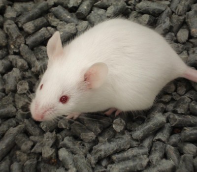
Animal Aid has uncovered extremely disturbing experiments involving live mice being surgically joined together, and forced to remain like this for several weeks. The research paper reporting these terrible experiments explains how the research was ‘supported in part by grants’ from various organisations, including the UK Multiple Sclerosis Society1. A separate research paper involved a toxin being injected into the brains of mice in order to damage them. The mice were later killed and their brains analysed. Two researchers listed on this paper were funded by the UK Multiple Sclerosis Society2; one of them co-authored the paper about the experiments that involved mice being stitched together.
Animal Suffering
- As well as the physical effects, such as pain or infection, which can arise from being surgically attached to another animal, there would also undoubtedly be mental distress from this horrific procedure. Anyone who has tried to run in a three-legged race, will know how frustrating, jarring and difficult it can be – even when you have volunteered to do it, know what to expect and can be ‘released’ at any time, none of which applies to these mice. The mice would have been anaesthetised as single animals and awoken from the anaesthetic attached to another animal. The animals cannot go where they like, explore, feed, drink or sleep, unless their ‘partner’ also moves in the same direction. The psychological stress the animals suffer is potentially enormous and inescapable.
- Parabiosis, the surgical joining of two animals, has been described in other papers. One says, ‘in 1-2 weeks parabiotic mice have the ability to ambulate normally on surgically paired fore- and hind-limbs.’ 5 This means that, for 1 to 2 weeks, the animals are not able to walk, or move about, normally. So the mice face 1 to 2 weeks of impaired mobility which could cause pain as well as mental distress.
- The same paper also states ‘To ensure firm support and prevent pain, the suture connecting the elbows and knees should surround the joints rather than passing through the tissue’.5 This shows just how major an undertaking the procedure is.
- It is obvious that the mice who had their brains damaged by toxins would also have suffered.
- In addition to the suffering from the procedures outlined above, animals suffer from the very fact that they are kept in laboratories due to issues such as noise, lights and the presence of humans.
The experiments
- The experiments where animals were surgically joined are not described in the paper itself. Instead it refers to another paper3, which refers to a further paper4 where basic information is found of how the surgery was conducted. It describes how a mouse is shaved down one side and then skin is cut from the animal’s shoulder down to their knee on the same side. Another mouse is shaved on the opposite side of their body and cut from the same points. The animals then have their front legs tied to each other and their back legs tied to each other, around the joint. The cut along their bodies is widened to create an open section of skin and these, on both animals, are either stitched or stapled together – the paper does not elaborate which method is used.
- Three weeks after the animals were joined together, one of them would receive an injection of a toxin into their spinal cord. The skin on the back of the animal’s neck is cut open until the mouse’s spinal cord is exposed and the toxin is injected into the spinal cord. The mice are killed at various points after this injection.
- The animals who were not sewn together, who received the injection of a toxin into their brains, in order to cause damage, were killed at various points (3, 10 and 21 days) after the injection. After their deaths, their brains were analysed.
Scientific critique
- According to the NHS, ‘Multiple sclerosis (MS) is a condition which can affect the brain and/or spinal cord, causing a wide range of potential symptoms, including problems with vision, arm or leg movement, sensation or balance.’ They explain further that ‘MS is an autoimmune condition. This is when something goes wrong with the immune system and it mistakenly attacks a healthy part of the body – in this case, the brain or spinal cord of the nervous system. In MS, the immune system attacks the layer that surrounds and protects the nerves, called the myelin sheath. This damages and scars the sheath, and potentially the underlying nerves, meaning that messages travelling along the nerves become slowed or disrupted.’9
- MS is described as ‘a uniquely human disease. It does not occur naturally in any other animal.’6 This is yet another example of artificially inducing disease in animals in an attempt to mimic what happens naturally in some humans.
- The exact cause of multiple sclerosis in humans is not clear – ‘the underlying cause may not be the same in all patients’ 6 – so the researchers are attempting, with toxic chemicals, to mimic something of unknown origin. The cause of demyelination in humans, is not an injection of toxin directly into the brain or spinal cord, which is what is being done in these cases.
- It has been pointed out in a research paper how the experimental ‘model’ differs from the human situation. Regarding demyelinating lesions (areas of damage to the myelin sheath): ‘It must be remembered that nearly all demyelinating lesions in experimental animals are small compared to demyelinating lesions in humans and most will undergo spontaneous remyelination’.7 Not only are mice different from us in many ways, but also the ‘model’ which is being used is different from the human conditions in the size of the damaged areas and the ability for these to be reversed by the body.
- A paper which describes mouse ‘models’ used in MS research states, regarding toxin-induced models of demyelination, which are used in these experiments state that they: ‘do not reproduce other aspects of MS, such as inflammation and axonal loss’.6 So some very important elements of the disease in people are not being reflected.
- When discussing mouse ‘models’ of MS one paper states: ‘the processes of demyelination and remyelination are less extensive and more acute than in the human condition’ 8 which means there are even more differences between how this disease naturally occurs in humans and how it is ‘modelled’ in mice.
- In the paper describing parabiosis, the researchers describe pairings of animals in one phase of the research. There are three treatment groups with three, two and four pairs of animals respectively, with no explanation of why the sample groups are different sizes. Additionally, if two pairs are all that is ‘needed’ in one group, then surely this should be sufficient for the two other groups, which would have spared three pairs of animals suffering the parabiosis.
Additional information and background notes
- The annual income of the MS Society, in their last annual report, is stated as being £27,743,000. https://www.mssociety.org.uk/sites/default/files/Annual%20Report%20and%20Accounts%202015.pdf – accessed 2/6/17
- The MS Society explains, in their 2015 Annual report, that their legacy income was £9.3 million (34% of their income).
- To find out more about how the public can unwittingly ‘Leave a legacy of suffering’: http://victimscharity.wpengine.com/legacy-of-suffering/
- To find out more about why ‘Mice Matter’ and they should not be used as ‘research tools’ in laboratories, visit www.micematter.org.uk
References
- Ruckh, J.M. et al (2012) ‘Rejuvenation of Regeneration in the Aging Central Nervous System’, Cell Stem Cell, vol.10, pp. 96-103.
- Miron, V.E. et al (2013) ‘M2 microglia/macrophages drive oligodendrocyte differentiation during CNS remyelination’, Nature Neuroscience, vol. 16, no.9, pp.1211-1218
- Wagers et al (2002) ‘Little evidence for developmental plasticity of adult hematopoietic stem cells’, Science, vol.297, pp.2256-2259
- Wright, D.E. et al (2001), ‘Physiological Migration of Hematopoietic Stem and Progenitor Cells’, vol.294, issue. 5548, pp.1933-1936.
- Kamran, P et al (2013) ‘Parabiosis in Mice: A Detailed Protocol’, Journal of Visualized Experiments, vol.80, e50556.
- Pachner, A.R. (2011) ‘Experimental models of multiple sclerosis’, Current Opinion in Neurology, vol.24, pp.291-299
- Blakemore, W.F. & Franklin, R.J.M. (2002) in M. Rodriguez (eds) ‘Advances in Multiple Sclerosis and Experimental Demyelinating Diseases, Current Topics in Microbiology and Immunology, pp.193 – 212
- Miller, R.H. et al (2010) ‘Restoring the balance between disease and repair in multiple sclerosis: insights from mouse models’, Disease Models & Mechanisms, vol.3, pp.535-539
- ‘Multiple sclerosis’ – http://www.nhs.uk/conditions/Multiple-sclerosis/Pages/Introduction.aspx – accessed 6/6/17